
What is Ossicular Chain Reconstruction?
Ossicular chain reconstruction (OCR), or ossiculoplasty, is a specialized surgical procedure aimed at repairing or replacing the small bones of the middle ear—malleus, incus, and stapes—that are essential for hearing. Damage or interruption in this ossicular chain can significantly impair the conduction of sound from the eardrum to the inner ear, resulting in conductive hearing loss.
OCR is often performed in conjunction with other otologic surgeries, such as tympanoplasty (repair of the eardrum) or mastoidectomy (removal of infected mastoid bone). The primary objective is to re-establish a functional mechanical pathway for sound vibrations to reach the cochlea, thereby improving the patient’s hearing capacity.
Anatomy and Physiology Review: Middle Ear and Ossicular Chain Function
The middle ear is an air-filled space behind the eardrum (tympanic membrane) that houses three tiny bones known as the ossicles. The three ossicles are connected by joints that move, just like any other bone in the body. In order, from eardrum to inner ear, the ossicles are:
-
1. Malleus (hammer): Attached to the eardrum and acts as the initial receiver of sound vibrations.
-
2. Incus (anvil): Serves as a bridge between the malleus and stapes.
-
3. Stapes (stirrup): The smallest bone in the human body, transmits sound vibrations to the oval window of the cochlea in the inner ear.
These bones form a lever-like mechanism that amplifies the sound that hits the eardrum by a factor of 22! This amounts to about 25-30 dB of sound amplification. It is therefore easy to understand why this energy transformation from mechanical-to-hydraulic, or air-to-fluid, (from the outer ear which contains air to the inner ear which contains fluid) is vital for efficient hearing.
When the ossicular chain is disrupted due to disease, trauma, or congenital conditions, the ossicles' amplification system is compromised, leading to a reduction or loss of sound conduction.
What Materials Are Used For Ossicular Reconstruction?
Ossicular reconstruction is a surgical technique designed to restore the continuity of the middle ear’s tiny bones by connecting gaps or replacing sections that have been damaged or lost. Doing so will provide a stable and properly aligned mechanical path that transmits vibrations from the eardrum to the oval window (inner ear), ensuring optimal hearing restoration. To achieve this, surgeons use three main approaches:
1. Surgical prostheses (implants):
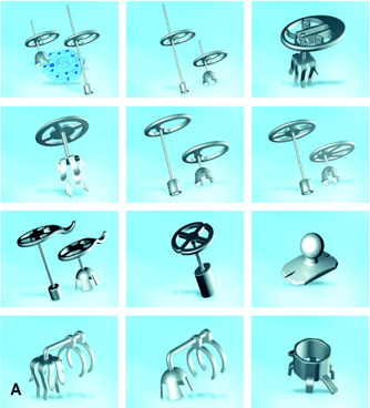
Prostheses (or implants) are man-made from biocompatible materials like titanium, Teflon and hydroxyapatite (a form of ceramic that mimics bone). They are precisely shaped to mimic the natural ossicles and reestablish contact between the eardrum and the inner ear. Prostheses come in a variety of shapes and designs, each tailored to address specific anatomical and surgical needs. During the procedure, the surgeon will carefully select the type and length of prosthesis that best matches his patient’s unique ear structure and the extent of ossicular damage. This customization is essential to ensure the most effective restoration of hearing and achieve optimal surgical outcomes. The two most common prosthesis I use are PORPs (partial ossiculaire replacement prosthesis) and TORPs (total ossicular replacement prosthesis). The image below shows different types and shapes of protheses.
2. Bone cement (Otomimix):
Otomimix is a bone cement made of hydroxyapatite (a form of calcium mineral). As stated in the paragraph above, hydroxyapatite mimics the consistency of human bones. During surgery, the bone cement is first prepared as a soft, moldable paste. The surgeon carefully shapes this paste to fill the exact area of bone loss or damage in the middle ear. After it is positioned, the cement is allowed to set for a few minutes. Once hardened, it closely resembles the density and properties of the natural ossicle, effectively restoring the missing connection needed for sound transmission.

3. Autologous grafts:
Autologous means "from your own body". Therefore, these grafts involve using the patient’s own ossicles (if they are spared by the infection or other diseases and can be re-used safely) and reshape them to bridge the gap between the eardrum and the inner ear (or remaining ossicles). The most common type is called "incus interposition graft", where the patient's remaining incus will be used to connect the malleus to the stapes or inner ear. This is only possible if the incus and residual ossicles are healthy enough to be used, which is not common in patients with chronic ear diseases and infections. These reconstructions integrate well and reduce the risk of rejection that can be seen with implants.
When to Do Ossicular Chain Reconstruction (Indications)
OCR is indicated in a variety of clinical scenarios where the ossicular chain is disrupted or malformed. Common indications include:
-
Chronic otitis media: Repeated middle ear infections can erode the ossicles, particularly the long process of the incus.
-
Cholesteatoma: A skin cyst that grows in the middle ear and mastoid, destroying surrounding structures, including the ossicles.
-
Trauma: Physical injury, including skull base fractures or barotrauma from rapid pressure changes, may dislocate or fracture the ossicles.
-
Congenital anomalies: Some individuals are born with malformed or missing ossicles, resulting in hearing loss from birth.
-
Revision surgery: Patients who have previously undergone ear surgery and have persistent or recurrent conductive hearing loss due to ossicular chain damage or scarring.
Types of Ossicular Reconstruction Procedures
A. Incudo-Stapedial (IS) Joint Reconstruction
The incudo-stapedial joint (the joint between the last two bones, the incus and the stapes) is a frequent site of erosion. The type of the surgical correction depends on the how bad the damage is:
-
Otomimix (bone cement): Used for small erosions where a fine gap exists between the incus and stapes. The cement is molded into the desired shape to reconnect the joint. It hardens quickly and provides a solid bond that fixes the continuity of the joint.
-
Incus interposition graft: If the incus is too damaged to use Otomimix, it is entirely removed and re-shaped to fit the gap between the stapes and the malleus. Doing so will re-establish the connection between the inner ear and the eardrum. This is an "autologous" graft. Alternatively, a PORP can be used (see below).
B. Partial Ossicular Replacement Prosthesis (PORP)
PORPs are among the most commonly used implants to reconstruct the ossicles. They are typically made from biocompatible materials such as titanium or hydroxyapatite. A PORP is specifically used when the stapes bone (the innermost bone connected to the inner ear) is intact and mobile, but the malleus or incus bones (the outer and middle ossicles) are damaged or missing. The prosthesis bridges the gap between the eardrum (or malleus) and the head of the stapes, restoring the transmission of sound vibrations to the inner ear. They are often used in cases of chronic ear infections, cholesteatoma, or ossicular erosion due to trauma.
C. Total Ossicular Replacement Prosthesis (TORP)
A TORP is an also a very common type of implant used during ear surgery when all three ossicles—including the stapes—are damaged or absent, leaving only the footplate of the stapes in place (the base of the stapes). In this situation, the TORP acts as a complete bridge from the eardrum directly to the oval window of the cochlea (inner ear). It is longer than a PORP and must be carefully positioned to transmit sound vibrations effectively without damaging the delicate inner ear structures. Because it lacks the natural stapes anchoring point, it is more susceptible to displacement. TORPs are often indicated in severe cases of ossicular erosion due to chronic ear infections, cholesteatoma, or revision surgeries
Outcomes and Prognostic Factors (What affects the result)
Hearing improvement after OCR can vary widely based on the underlying condition, surgical technique, and individual patient factors.
Expected outcomes:
-
Most patients experience an average hearing gain of 20–30 decibels. This is a significant improvement no matter how bad the hearing was before the surgery.
-
Success rates range from 60% to 90% for closing the air-bone gap to within 10–20 dB (essentially fixing the mechanical part of the hearing loss to near-normal levels). Read this article to learn more about air-bone gaps.
What affects the outcomes of the ossiculoplasty? (prognostic factors):
-
Presence of a mobile stapes: The presence of a healthy and mobile stapes is the most important factor for the stability and sound transmission of the reconstruction (no matter the type).
-
Middle ear aeration: Good Eustachian tube function helps maintain middle ear pressure balance and prevents fluid buildup. It also prevents the eardrum from collapsing or retracting after surgery. This is also a very important factor. Read more about Eustachian tube function here.
-
Healthy middle ear mucosa: Chronic inflammation, granulation tissue and repeated ear infections can compromise prosthesis function.
- Number of ear surgeries: Repeated surgeries on the ear can reduce the chances of restoring good hearing, as each procedure increases scar tissue and decreases healthy blood supply.
-
Proper prosthesis positioning: Misalignment or prosthesis migration can limit hearing gain or cause extrusion.
-
Surgical field visibility: Better visualization through the canal or via a postauricular approach (behind the ear) allows for more accurate placement.
- Smoking: Smoking significantly lowers the chances of successful eardrum healing after surgery, which can directly impact how well the ossiculoplasty recovers.
- Patient medical factors: Patients with diabetes, weakened immune systems, or a history of head radiation may experience slower or less complete healing after surgery.
What to Expect After OCR Surgery
OCR surgery is often combined with a tympanoplasty or a mastoidectomy. For the recovery following those two surgeries, please read the relevant articles (see this article for tympanoplasty).
This section below will deal specifically with OCR (done through the ear canal).
Immediate post-operative period:
-
Pain is generally mild and controlled with over-the-counter medications.
-
A sensation of ear fullness or mild imbalance is common for a few days.
-
The ear is typically packed with a special absorbable dressing to support grafts and prostheses.
Recovery timeline:
-
The outer ear dressing and sutures (if used) are removed within 1–2 weeks.
-
Most patients return to daily activities after 1 week, avoiding strenuous activity, nose blowing, or water exposure.
-
Audiologic testing is performed 4-6 months after surgery to assess hearing outcomes.
Precautions:
-
Avoid getting water in the ear until cleared by your surgeon.
-
Refrain from flying or rapid altitude changes for 4–6 weeks.
- Avoid heavy physical activity (less than 15 lbs), work-outs, swimming, etc. for a period of 6-8 weeks.
-
Do not use Q-tips or insert anything in the ear canal.
Follow-up care:
-
Patients are monitored for signs of infection or prosthesis displacement with routine clinic visits.
-
A repeat audiogram will be done at 1 year after surgery.
-
In some cases, revision surgery may be necessary if the initial reconstruction fails.
Conclusion
Ossicular chain reconstruction is a highly effective surgical solution for conductive hearing loss due to ossicular damage. With proper assessment, surgical planning, and post-operative care, most patients experience significant improvement in their hearing and quality of life. If you have chronic ear disease or hearing loss suspected to involve the middle ear bones, consult with an otologic surgeon to determine if OCR could benefit you.

Joe Saliba, MD
Dr. Joe Saliba is an ENT surgeon specialized in neuro-otology and medical director at ODYO. He treats patients with various ear and skull base disorders, ranging from hearing loss and vertigo to vestibular schwannomas and cochlear implants.